
Lymphatic filariasis is a parasitic disease that is transmitted to humans, like malaria and yellow fever, via a mosquito bite. However, unlike those acute infections, lymphatic filariasis may not manifest itself for years or decades after the initial infection.

Image/CDC
Though there are millions of people infected throughout the world, this parasitic disease hasn’t been seen in the U.S. in about 100 years.
There are three species of parasites that cause lymphatic filariasis–Wuchereria bancrofti; which is more widely distributed; Asia, Africa, India, South America and some Caribbean Islands and Brugia malayi and B. timori which are more restricted to parts of Asia.
These parasites are transmitted by several species of mosquito; Culex, Anopheles, Aedes and Mansonia depending on the geographic area.
When the mosquito takes a blood meal on a person, it injects parasitic larvae onto the skin, where it penetrates the bite wound.
After which in time the larvae develop into adults (females can be up to 100 mm in length) and reside in the lymphatic system of the upper or lower limbs or groin (all species). With W. bancrofti, in human males the adult worms may end up in the lymphatic channels of the spermatic cord.
Here the adult male and female worms mate and produce eggs (microfilariae) which circulate in the blood and lymph. The microfilariae only appears in blood at certain times; Wuchereria at night, Brugia during the day.
Most infections are asymptomatic. Any disease present may be due to immune response. If the infection persists the chronic stages of disease develop.
It will then go into an inflammatory stage where lymphadema, orchitis and hydrocele occur.
The obstructive stage of the disease is called elephantiasis. In this stage, which may take years, there is a blockage of lymph flow due to masses of worms. Tissue becomes fibrotic and skin thickens.
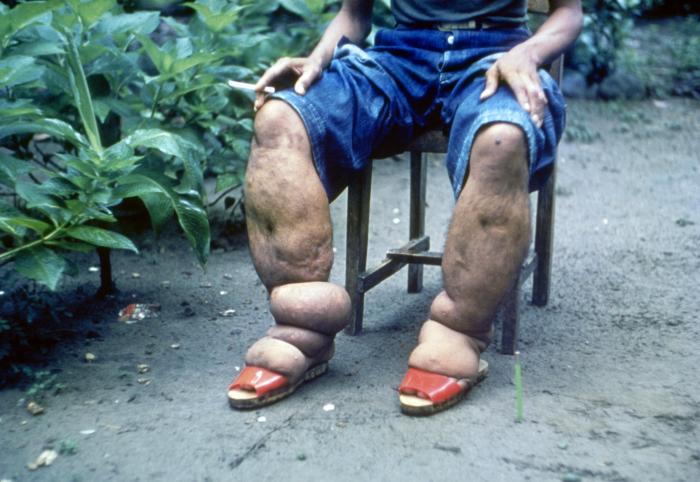
Enlarged legs, arms, mammory glands and genitalia are classic appearances of elephantiasis.
Diagnosis in the acute stages can be made by finding microfilariae in blood smears. Antigen detection and molecular methods can also be used to diagnose. Microfilariae are not found in persons with elephantiasis.
Diethylcarbamazine (DEC) is the drug of choice. The drug kills the microfilaria and some of the adult worms.
Later stages of the disease require different treatment. According to the Centers for Disease Control and Prevention (CDC), lymphedema and elephantiasis are not indications for DEC treatment because most people with lymphedema are not actively infected with the filarial parasite.
To prevent the lymphedema from getting worse, patients should ask their physician for a referral to a lymphedema therapist so they can be informed about some basic principles of care such as hygiene, exercise and treatment of wounds.
Patients with hydrocele may have evidence of active infection, but typically do not improve clinically following treatment with DEC. The treatment for hydrocele is surgery.
There is not a vaccine to prevent filariasis. Travelers to endemic areas should use mosquito repellent on exposed skin between dusk and dawn.
For more infectious disease news and information, visit and “like” the Infectious Disease News Facebook page
